One project keeps chemist Wei Kong awake at night, and it started as an idea nearly two decades ago. Now, after being awarded nearly $2 million for four years by the National Institutes of Health, the goal is to create a groundbreaking new tool with the potential to revolutionize drug development and enhance our understanding of disease mechanisms.
At the core of this project lies the significance of molecular structures. Unraveling the intricacies of molecules is crucial for discovering new drugs and understanding the formation and progression of diseases.
While there are existing tools in the field currently, they all have limitations, including crystallography and cryo-electron microscopy. There is also a pressing need for equipment to work on molecules of different sizes and purities, an unmet challenge currently.
“We have a whole new playground to investigate materials,” Kong said. “We are the first one in the whole world who can do diffraction in a droplet. And the structures formed in the droplet can be different from the structures formed in any other environment.”
A new scientific frontier
Serial Single molecule Electron Diffraction Imaging combines multiple tools. The process starts by pulling proteins into a vacuum using electrospray ionization, a technology pioneered by chemist John Fenn, who was awarded the Nobel Prize for its development. Electrospray ionization generates intact gas-phase ions for mass spectrometry.
Simultaneously, high-pressure pre-cooled helium is sprayed into a high vacuum, forming superfluid helium droplets with an ultralow temperature of 0.4 Kelvin, equivalent to an incredibly frigid -459 degrees Fahrenheit. The resulting droplets exhibit zero friction, hence an embedded molecule inside the droplet can rotate freely.
Because the droplets capture anything along their path, they end up pulling in the proteins and cooling them to the same temperature, preventing denaturing and slowing down their rotation. Due to the repellent nature of identical charges, each droplet only contains one protein.
Next, the droplets are oriented using an electric field and aligned using a laser field. By orienting a large number of proteins the same way, the researchers will be able to accumulate signals and decrease “noise” or random events.
The technology works in pulses, sending a sequence of proteins through the orientation and diffraction chamber at a rate of 10 times per second. After a few hours, the result is a very high-quality diffraction pattern; however, the work doesn’t stop there.
To create a three-dimensional picture, the polarization of the laser is rotated. The projections from all the different angles are combined to solve the structure, such as the connectivity of atoms, the type of bonds between them and the overall shape of the molecule.
“It’s been a long time in the making. It’s quite challenging,” Kong said. “Every technique has been demonstrated one way or another, but the final product has never been done.”
The project requires the team to address another new frontier—the characterization method of laser alignment of large macromolecules. People have demonstrated alignment with small molecules, but not big ones. The reason is the lack of a suitable method to determine the effectiveness of laser alignment of large molecules.
"...there’s a lot of new science that we’re discovering."
For small molecules, the technique of characterization involves a Coulomb explosion. Typically this requires two lasers: the first aligns the molecule, and the second explodes the molecules into ionized atomic fragments. The top goes one way and the bottom goes the opposite. When one captures an image of the top, the bottom is not detectable due to the precise alignment.
With only a couple of atoms, this is easy to do. But if one has a large molecule with tens of carbons going in the same direction, the picture gets messy.
As the project has developed, unexpected discoveries have also been exciting.
“This looks like an engineering project, but there’s a lot of new science that we’re discovering. A halogen bond of iodine was first observed in the gas phase in our system. The superfluid environment afforded by the droplets is opening doors to many new bonding and structure motifs of neutral and ionic clusters. So that’s a lot of fun doing that,” Kong said.
Conversely, the group faces many challenges including electrical problems, dust, cooling water and changes in team members.
Even with protections in place, there is no way to protect the heat-and-shrinking properties of the metal material, which affects the stability and reproducibility of the experimental apparatus. Power outages mean the system warms up and needs to be re-cooled and then reconditioned for settlement.
Dust is a problem because it can accumulate on the detector and will eventually cause black spots on the screen and affect the data. And every time students graduate, it takes substantial time to train the next group on safety and equipment management.
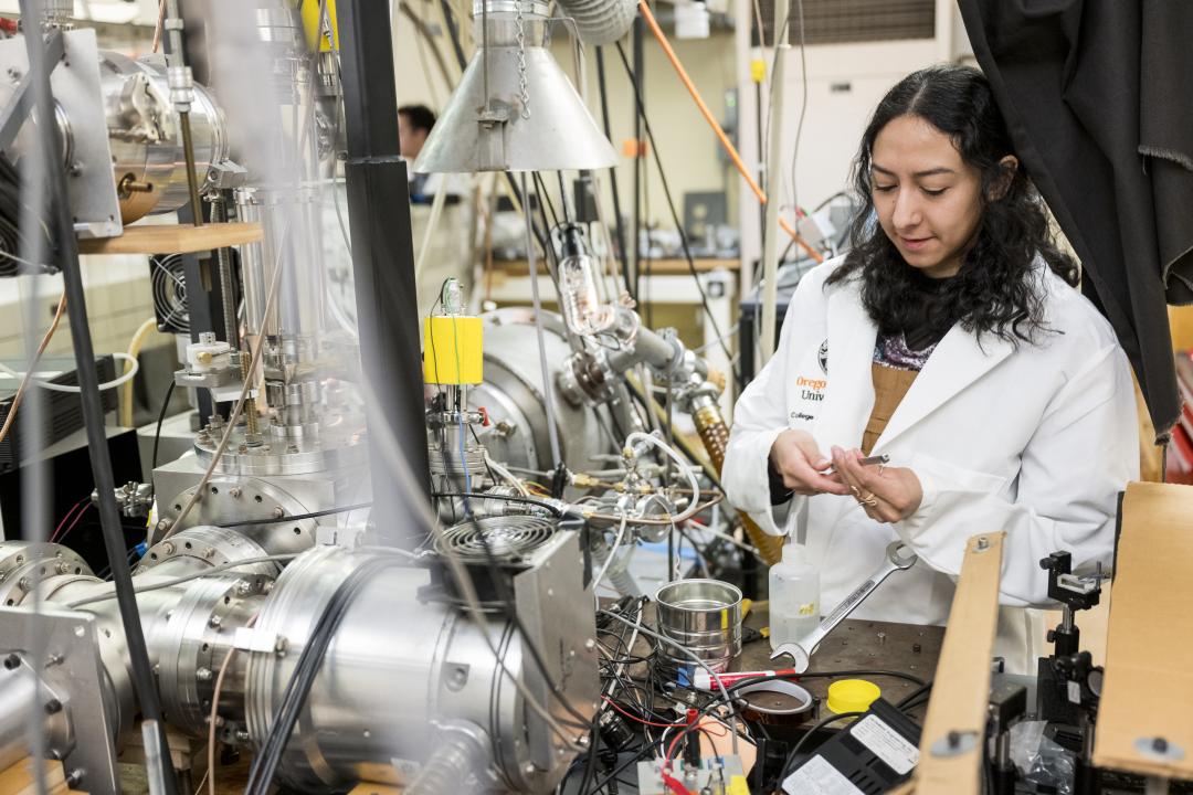
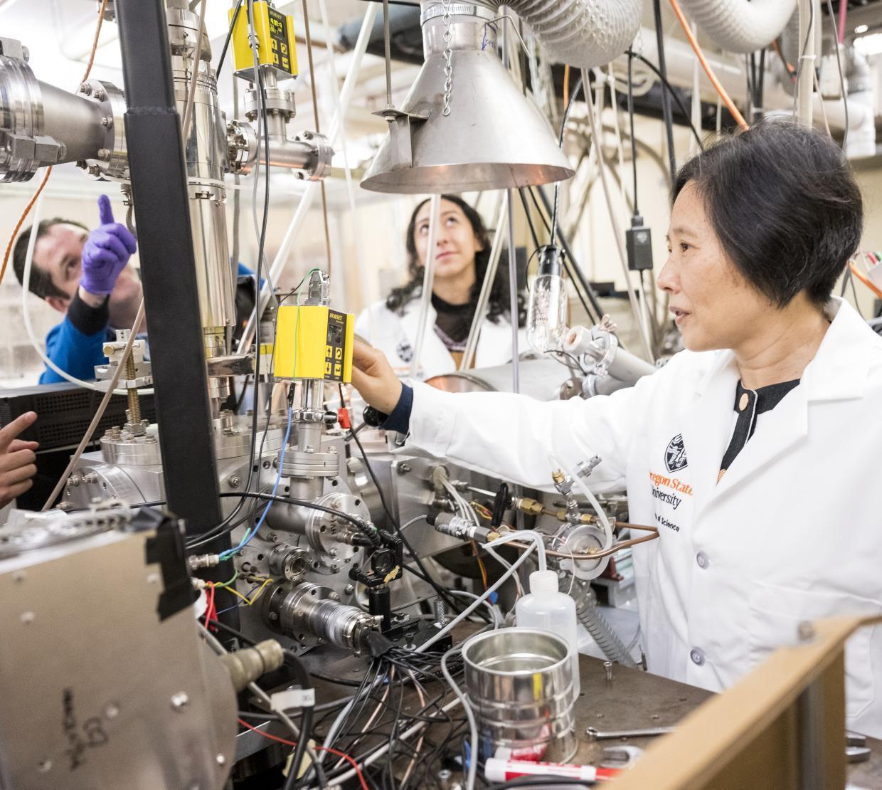
Presently, three graduate students—Marisol Trejo, Andrew Clifford and Joachim Schuder—are working with Kong on this project.
“They are all great— a dream team,” Kong said. “Even though we’ve had two or three generations of students that have not reached the final goal, every step, every milestone was satisfying and rewarding. That’s what keeps everybody going.”
Despite the challenges encountered along the way, this project marks a significant technological advancement and emphasizes the importance of perseverance and collaborative efforts in science.